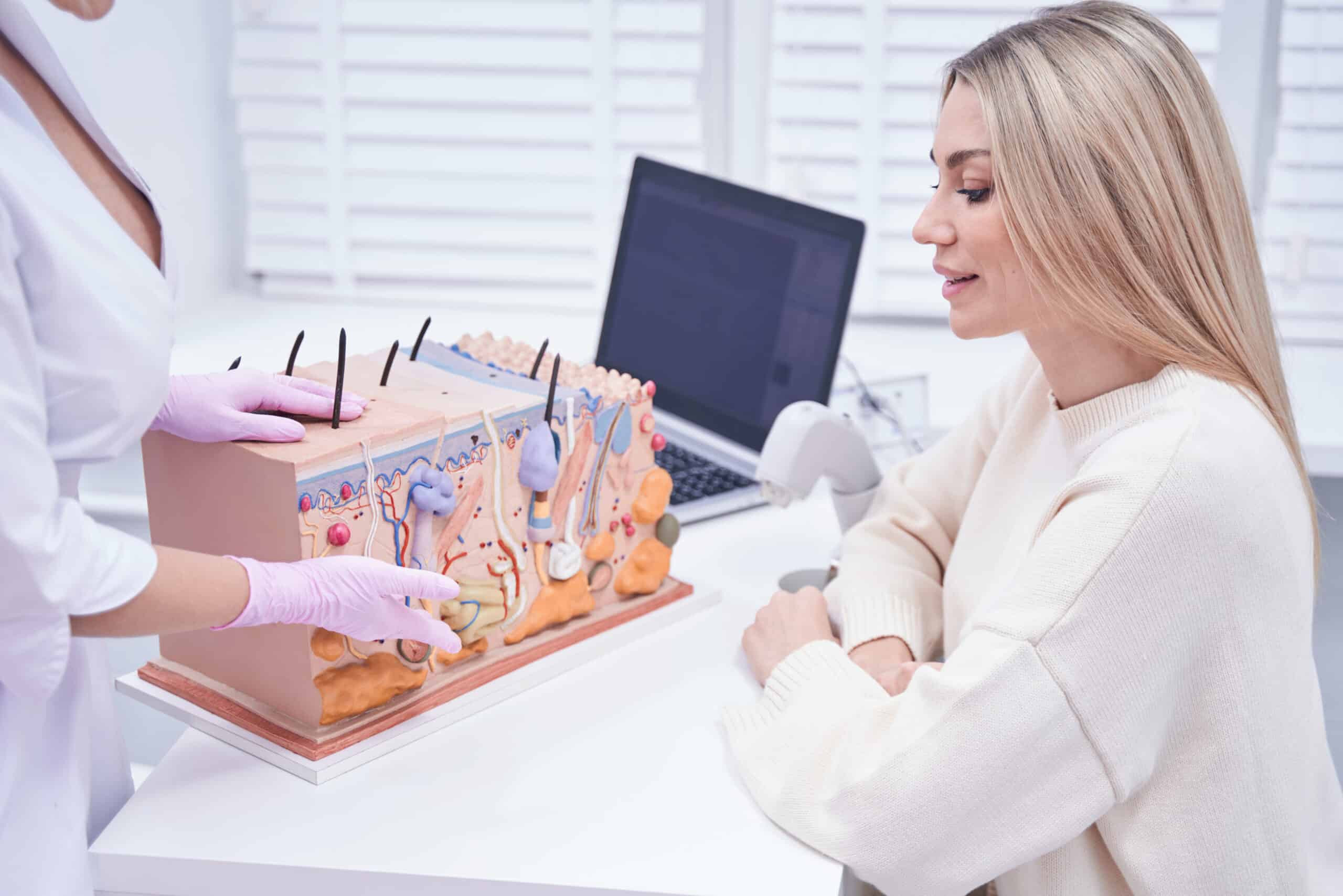
Our skin is comprised mainly of three layers: the epidermis (the top layer), the dermis (which lies beneath the epidermis), and fat (which lies beneath the dermis) (Abdo et al, 2020). The skin is mainly responsible for: protection against foreign bacteria/pathogens, maintaining body temperature and hydration, and the creation of Vitamin D. (Abdo et al, 2020).
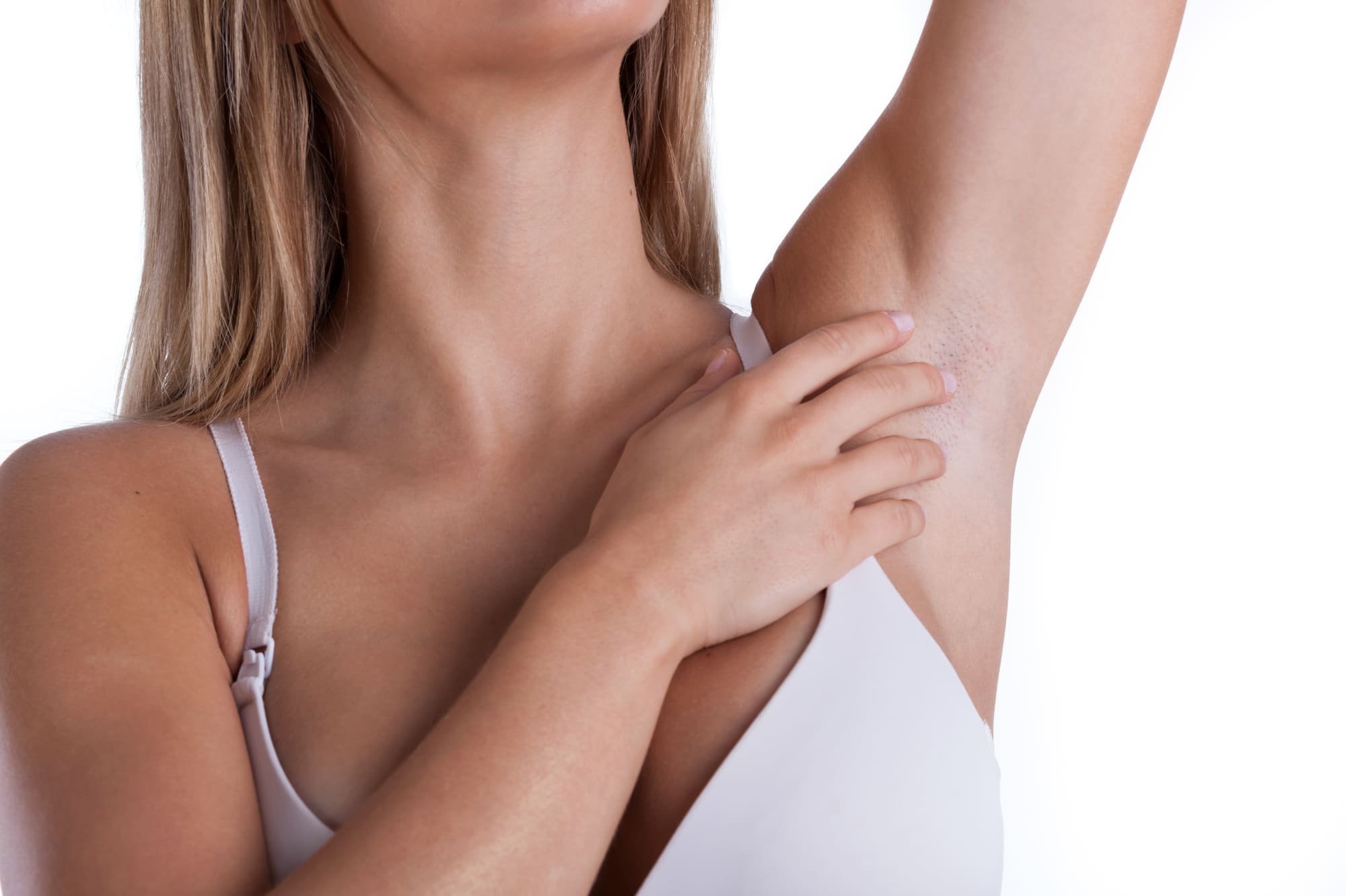
The epidermis is the outermost layer of the skin. The epidermis creates melanin, which gives us the color of our hair, skin, and nails (Abdo et al, 2020). The epidermis also maintains keratinocytes, which are cells that help with wound healing, transportation of water, immunity, and aids in melanin transport (Abdo et al, 2020). Cells in this layer are consistently turning over, which occurs every 28 days depending on skin hydration and age (Abdo et al, 2020). When undergoing laser treatments, typically the melanin found within brown spots is the target. These cells are found in the epidermis. One thing that is important to note is that if exposed to the sun, the body increases the activity of the melanocytes. This causes the laser/IPL to accidentally target the activated melanocytes versus the cells found in the brown spots and it can cause delayed healing. This is the main reason why avoiding the sun for a minimum of 2 weeks pre-treatment and 2 weeks post-treatment is so crucial. It is important to note that with certain treatments, for example, Micro-needling or Halo treatments, the epidermis is disrupted. Therefore, gentle skin care is essential for about 4-7 days post-treatment or until healing is complete. To prolong your results post-treatment, it is important to use sunscreen frequently and reapply generously. UV light exposure will cause more brown spots and continue to damage your collagen, so it is crucial to keep up with your SPF regimen.
The dermis is the layer of skin located beneath the epidermis. The dermis contains our blood vessels, collagen and elastin, and nerves (Hunter, 1973). One reason pinpoint bleeding seen after Microneedling is considered a positive outcome is that injury to the dermis generates collagen. Bleeding is a positive sign that practitioners have targeted the appropriate layer of the skin. Using lasers and/or Microneedling systems, we can effectively stimulate collagen creation by using appropriate settings.
References:
Abdo, J., Sopko, N., & Milner, S. (2020). The applied anatomy of human skin: A model for regeneration. Wound Medicine, 28. Https://doi.org/10.1016/j.wndm.2020
Hunter J. A. (1973). Diseases of the skin. Structure and function of the skin in relation to therapy. British medical journal, 4(5888), 340–342. https://doi.org/10.1136/bmj.4.5888.340